FACILITIES
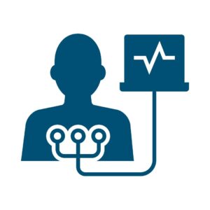
Electrocardiogram
(ECG)
OVERVIEW
- An electronic device which records the electrical signals produced by the heart muscle and produces electrocardiograms (abbreviated as ECG or EKG). ECGs are collected non-invasively and the procedure involves no pain. An ECG is a common and very effective test in order to understand the heart’s health.
- Though ECGs at the clinic possess better accuracy, they can now be taken at home using a smartwatch.
OBJECTIVE
- An ECG test is performed to determine if there are any heart problems and to understand heart’s health. An ECG can be used to determine if the patient is having or had a heart attack, if there is a blockage in the artery or if the heartbeats are irregular.
- In most cases, an ECG test is performed when a patient has reported symptoms of a heart issue such as breathlessness, chest pain, cold sweating, increased heartbeats and lightheadedness. The vital data collected during the test is then inspected by a doctor to determine further tests or treatments.
PROCEDURE
- The objective of the test is to collect electrical signals generated by heart.
- For the detection, ten different leads or electrodes are placed on the patient’s body. These are divided into two types: four limb leads and six chest leads. The places where the leads are to be placed will be cleaned before the placement.
- The patient will feel no pain and will need to hold still for some time for accurate ECG readings. The collected tracings are printed on paper to be inspected by the doctor.
2D Echo &
Colour Doppler
OVERVIEW
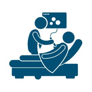
- A 2D echo test is performed to scan the heart muscle using high-frequency ultrasound waves. When combined with the colour doppler technique, which uses the frequency of the sound waves, it provides important data such as blood flow, the pressure of the flow across different regions of heart and if there are any obstructions in the blood flow.
- The test is non-invasive, painless and has no side effects.
OBJECTIVE
- The test is performed to determine if there are any structural or functional defects in the heart muscle. Pumping functionality of the heart, wall motion of the heart muscle, functioning of the heart valves are checked along with dimensions of the chambers, abnormalities of the heart & structure of the valves.
- The test is performed if the patient has reported heart-related issues such as breathlessness, chest pain, cold sweating, increased heartbeats, and lightheadedness.
PROCEDURE
- The test is non-invasive and is painless.
- The patient would be asked to uncover the chest area and to lie down. A gel will be applied to the chest and the doctor will move a transducer over the area to view the heart muscle and other structures.
- A transducer is a small remote-like device that is used to send the signals through the area. After the images are collected, the gel is then removed and the reports will be delivered.
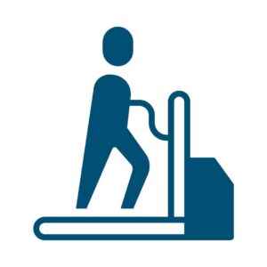
Stress Test
(TMT)
OVERVIEW
- An exercise stress test or a treadmill test is aimed towards determining if heart receives enough oxygen and blood flow when it needs it the most, such as when the patient is exercising.
- The test is simple, non-invasive and involves no pain during the procedure but the patient might feel a little exhausted due to the exercise involved. The test helps to guide the treatment decisions made for the patient.
OBJECTIVE
- A stress test is recommended if the patient reports having heart-related issues such as heaviness in the chest, fatigue, lightheadedness, breathlessness with or without physical activity and rapid heartbeats.
- The test is advised if the doctor suspects that the patient has coronary artery disease or an irregular heart rhythm, or just to ensure the overall health of the heart muscle.
PROCEDURE
- The test requires the patient not to consume food and to smoke for some time before the test. A doctor would ask different questions that might affect the result of the test and decide on the right amount of exercise as per the patient’s physical condition.
- Before the test, a blood pressure monitoring pad would be placed on the patient’s arm while some ECG leads or electrodes would be placed on the chest, legs and arms as needed. During the test, the patient would be asked to step on a treadmill and start walking slowly. As time passes, the speed would increase and the data would be collected to check how the heart acts to cope with the stress buildup.
- The exercise would continue till the target level is reached or the patient shows symptoms that don’t allow them to continue the test, such as breathlessness, fatigue, chest pain, lightheadedness, etc.

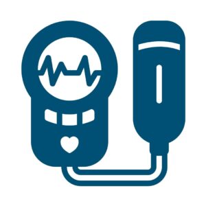
24 Hour Ambulatory
BP Monitoring
OVERVIEW
- A 24 hour ambulatory blood pressure monitoring is used to collect blood pressure & heart rate reading over the period of 24 hours or more.
- These readings are then averaged over the 24-hour period. Important data such as average heart rate, changes in the heart rate, and distribution of the blood pressure over 24 hours can be understood by the doctor to manage or monitor hypertension.
OBJECTIVE
- 24 hour ambulatory blood pressure monitoring is recommended for patients who suffer from white coat hypertension, which is having elevated blood pressure in the healthcare setting or for patients who suffer from masked hypertension, which is having lower blood pressure at the doctor’s office but elevated blood pressure at home.
- The test is also advised for pregnant women with hypertension, people with borderline hypertension, people with hypotension, and people having difficulties controlling blood pressure using medications.
PROCEDURE
- The test is non-invasive and involves no pain for the patient.
- The doctor will provide a device and this device will be placed over the belt or strap worn over the body of the patient. The device would have a cuff attached to it, which would be wrapped around the upper arm of the patient. The cuff would inflate at certain intervals throughout the day and night to record the blood pressure.
- These readings would be collected or the patient would be asked to keep a record.

24 Hour Holter
Monitoring
OVERVIEW
- If the ECG of a patient does not provide the doctor with sufficient data for treatment, then 24-hour holter monitoring is recommended. This will be advised only if the ECG does not provide enough details of the patient’s heart health.
- A Holter monitor is a small device that records the heart rhythm of a patient. The device can be worn by the patient inside of their clothing.
OBJECTIVE
- The test is advised after a conventional test such as ECG couldn’t provide enough data for the assessment of the patient’s problems.
- A Holter monitor test is performed if the doctor suspects an irregular beating pattern of the patient’s heart, if the patient has unexplained fainting or if there is a chance of irregular heart beats that ECG missed.
PROCEDURE
- The holter monitor device will be provided to the patient by the doctor at the office. The device should be worn constantly on the body for the described time.
- As the device should not come in contact with water and should not be detached, the patient might be asked to take a bath before visiting the doctor if the device is to be worn for more than a day.
- The electrodes, or the leads from the device, will be stuck to the body by the doctor, and the device will be attached by a strap to the body. The device is about the size of a deck of cards. After the placements, the patient can return to the usual activities and might need to record some details as advised by the doctor.
- The test is non-invasive and involves no pain for the patient.

Peripheral, Arterial &
Venous Doppler
OVERVIEW
- These Doppler tests are performed to understand the way the blood flows through the patient’s legs or their hands.
- Normal ultrasound provides images of the blood vessels but with the Doppler technique, the blood flow or blood circulation can be visualised along with any blockages that may be present in the blood vessels of the patient’s body.
OBJECTIVE
- The test is performed if the patient has issues such as leg pain, leg cramps or fatigue.
- There could be other complaints, such as slow healing ulcers with or without a family history of peripheral artery disease. If the doctor suspects any chances of blockages in the vessels, the test may be advised.
PROCEDURE
- These doppler tests are all non-invasive, can be easily done and involve no pain for the patient. There is no use of radiation, and hence the tests do not have any side effects either.
- The doctor would ask the patient to lie down on the examination table, after which a soluble gel would be applied to the areas to be inspected. The doctor would press the transducer firmly against the body and move it as needed to capture the required images and other data.
- The patient will not feel any pain during or after the procedure and can resume the usual activities after the completion of the test.
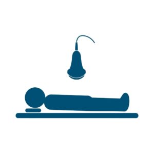
Abdominal
Sonography
OVERVIEW
- Abdominal sonography or abdominal ultrasound is performed to understand the condition of the structures inside the region. This test allows the doctor to view inside the abdominal area and the organs nearby to assess the issue that a patient might have, such as unexplained bloating and abdominal pain.
- The procedure is non invasive, uses low-power sound waves and is considered to have no side effects.
OBJECTIVE
- The test is performed if the patient carries the risk of having an abdominal aortic aneurysm. These risks involve a family history of the disease or smoking.
- This test may be recommended if the patient is considered to have issues in the kidneys, intestine, pancreas, gallbladder, liver, spleen or in the abdominal aorta, a major blood vessel that supplies blood to the entire body.
PROCEDURE
- The patient might be asked to avoid drinks and food for a specified time before the test, as this might not provide clear images of the internal structures. Upon arrival at the clinic, the patient would be asked to remove their clothing and to wear a hospital gown for the procedure.
- A soluble gel would be applied to the abdominal area and the radiologist would press a remote-like device called a transducer against the patient’s abdomin. This device would send signals to collect images of the structures inside the area.
- The patient would feel no pain & after completion of the test can resume the normal routine immediately.

Obstetrics
Sonography
OVERVIEW
- Obstetrics sonography is a non-invasive, painless test performed to check the condition of the fetus, ovaries, cervix and uterus of a pregnant woman.
- The test does not use any radiation and hence possesses no side effects for the mother or the baby inside of her.
- The procedure is very simple, provides a variety of important data, is widely available and is affordable.
OBJECTIVE
- The clinical test is performed to confirm the presence of a living foetus, evaluate the position of the foetus and check for abnormalities and to check the growth of the baby along with its overall health.
- The test also provides other vital data such as fluid around the baby, opening or shortening of the cervix, any abnormalities of the ovaries or uterus, etc.
PROCEDURE
- The procedure requires the radiologist to place a transducer on the exposed stomach, so the patient would be advised to wear comfortable clothes that can give the radiologist access to the area.
- During the test, a soluble gel will be applied to the patient’s stomach and a transducer will be placed firmly against the skin. This device would be moved as needed by the radiologist to take clear images of the internal structures for further inspection.
- Apart from a little pressure on the stomach area, the patient would feel no pain. The patient can resume their normal routine as soon as the test is over.
Renal
Doppler
OVERVIEW
- Renal Doppler uses ultrasound signals to determine the blood flow in and out of the kidneys of a patient.
- This test is non-invasive, uses sound waves with no side effects and is used to determine if there is any narrowing of the blood vessels or to check overall kidney health.
OBJECTIVE
- The test is recommended if the doctor suspects narrowing of the blood vessels leading to the kidneys.
- It is highly likely that a patient with uncontrolled high blood pressure and back pain has narrowed blood vessels.
PROCEDURE
- The patient would be asked to change into a hospital gown for better access to the abdomen area where the test would be carried out.
- A soluble gel would be applied to the abdominal area and a probe or transducer would be held up against the patient’s stomach to collect the signals. The patient might be asked to perform different breathing techniques and to roll over to one side to collect clear images of the organs.
- A mild to moderate pressure can be felt during the procedure as the radiologist moves the probe, but no sharp pain is involved during the procedure.

Carotid
Doppler
OVERVIEW
- Carotid doppler is an imaging test performed in order to examine the carotid arteries that are located in the neck region.
- The carotid doppler test reveals the speed and direction of the blood flow and related data to determine if there are any blockages in the artery obstructing the blood supply.
OBJECTIVE
- This test is performed if the doctor suspects plaque buildup in any of the arteries in the area restricting blood supply. The plaque buildup occurs due to coronary artery disease.
- The patient might report issues such as fatigue, lightheadedness, chest pain, etc.
PROCEDURE
- The test is non-invasive and uses sound waves of low power, posing no side effects.
- Upon arrival, the patient would be asked to lie down on the examination table or chair. A soluble gel would be applied to the area, and a transducer or probe would be placed against the neck area to collect the images. A patient would be asked to move the neck as needed, and a little pressure might be applied to collect clearer images.
- Apart from the pressure applied, the patient would feel no pain from the procedure.

Thyroid
Sonography
OVERVIEW
- Thyroid sonography is an imaging test that uses ultrasound signals to produce images of the thyroid gland in the neck region. The test is used to determine and confirm lumps and nodules suspected in the thyroid gland of the patient during a routine checkup.
- This test is non-invasive and uses sound waves that have no side effects.
OBJECTIVE
- The test is recommended if the doctor suspects a lump in the neck region. The test is performed to check whether the lump is arising from the thyroid or an adjacent structure.
- The test is also used to determine if there is presence of thyroid nodules, its growth and if it requires further treatment.
PROCEDURE
- The test is simple and can be carried out quickly.
- A soluble gel would be applied to the neck area and a probe or transducer would be held up against the patient’s neck region to collect the data. The patient might be asked to move their neck from side to side as needed for clear images.
- A mild to moderate pressure can be felt during the procedure as the radiologist moves the probe but no sharp pain is involved during the procedure.
Breast
Sonography
OVERVIEW
- Breast sonography is an imaging test performed in order to examine the internal structures of the breast region.
- The test is non-invasive, requires no special preparation and can be carried out without any side effects as the test uses ultrasound waves that are harmless.
OBJECTIVE
- This test is performed if the doctor suspects breast lumps or some other abnormalities found during the physical inspection.
- The doctor may perform a breast MRI or a mammogram before a breast sonogram is advised.
PROCEDURE
- The test is non-invasive and uses sound waves of low power, posing no side effects.
- Upon arrival, the patient would be asked to lie down on the examination table. The patient would be asked to expose the breast area while a soluble gel would be applied to the region under inspection. A probe or transducer would be placed on the area to collect the signals. This probe would be pressed firmly at times to collect clear images and would be moved to different locations for better angles.
- Upon completion of the test, the gel will be cleaned and the patient can resume regular work immediately.
WE
prioritise your queries.
prioritise your treatment.
prioritise your comfort.
prioritise YOU..!